Home

how to use To interact extremely false vocal cords radiology Colonel Against Headless

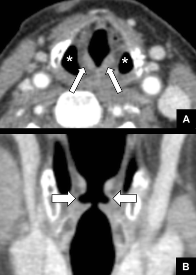
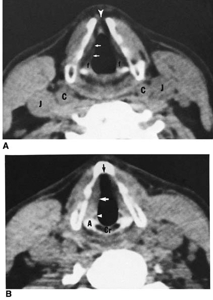
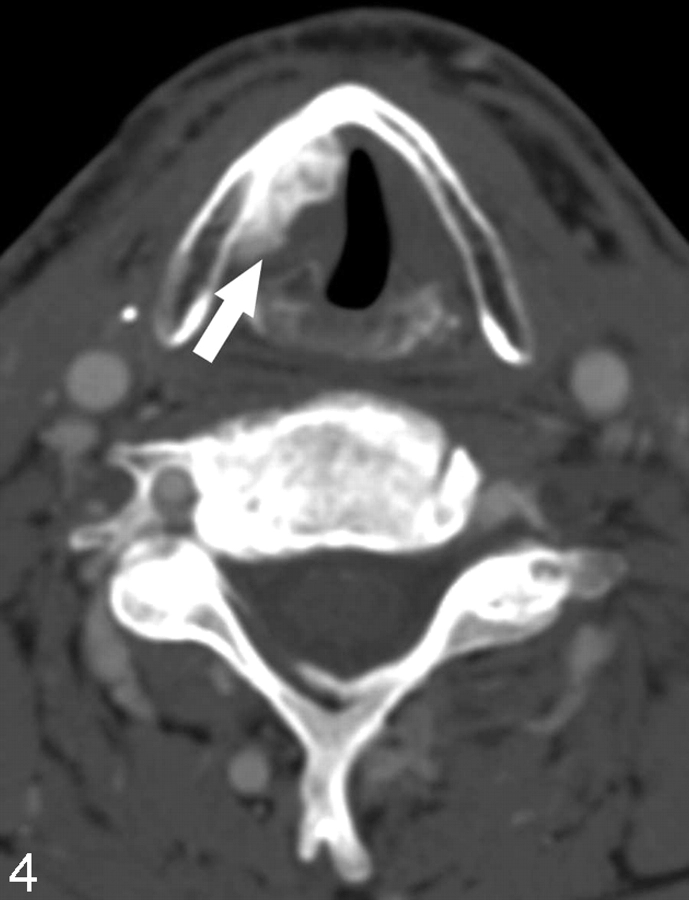
CT Assessment of Vocal Cord Medialization | American Journal of  Neuroradiology
CT Assessment of Vocal Cord Medialization | American Journal of Neuroradiology

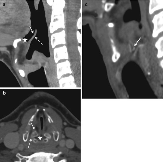
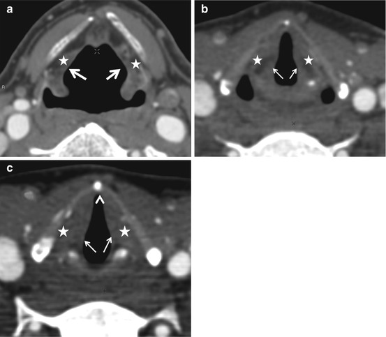
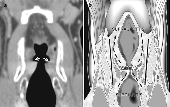
Vocal cord paralysis: anatomy, imaging and pathology | Insights into  Imaging | Full Text
Vocal cord paralysis: anatomy, imaging and pathology | Insights into Imaging | Full Text

Normal vocal cords on ultrasound | Radiology Case | Radiopaedia.org

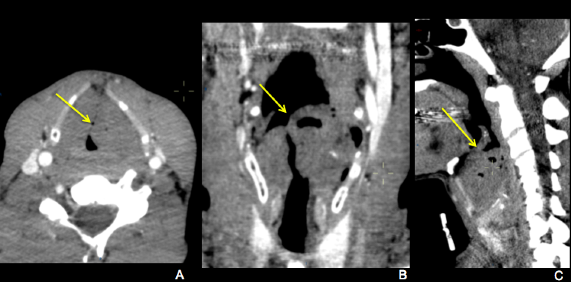
Axial and coronal cut sections of bilateral false vocal cords hypodense...  | Download Scientific Diagram
Axial and coronal cut sections of bilateral false vocal cords hypodense... | Download Scientific Diagram

Normal larynx. (a) Axial CT scan shows the normal appearance of the... |  Download Scientific Diagram
Normal larynx. (a) Axial CT scan shows the normal appearance of the... | Download Scientific Diagram

Normal Anatomy Mucosal Anatomy

Fiberoptic laryngoscopy shows a smooth bulge over left false vocal cord...  | Download Scientific Diagram
Fiberoptic laryngoscopy shows a smooth bulge over left false vocal cord... | Download Scientific Diagram

EPOS™
EPOS™

Vocal cord paralysis | Radiology Reference Article | Radiopaedia.org

Imaging of larynx - YouTube

Radiologic Evaluation of Larynx Cancer | Ento Key

Larynx and Hypopharynx | Radiology Key

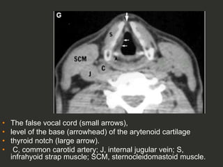
Normal anatomy of the false and true vocal cords on computed tomography

Radquarters - Supraglottic anatomy on axial CT. Supraglottic larynx  includes epiglottis, vallecula, aryepiglottic folds, false vocal cords, and  arytenoids. #FOAMrad #FOAMed #radiology #radres #radtech #medstudent  #neurorad #radiologisthq | Facebook
Radquarters - Supraglottic anatomy on axial CT. Supraglottic larynx includes epiglottis, vallecula, aryepiglottic folds, false vocal cords, and arytenoids. #FOAMrad #FOAMed #radiology #radres #radtech #medstudent #neurorad #radiologisthq | Facebook

Larynx and Hypopharynx | Radiology Key

CT Assessment of Vocal Cord Medialization | American Journal of  Neuroradiology
CT Assessment of Vocal Cord Medialization | American Journal of Neuroradiology

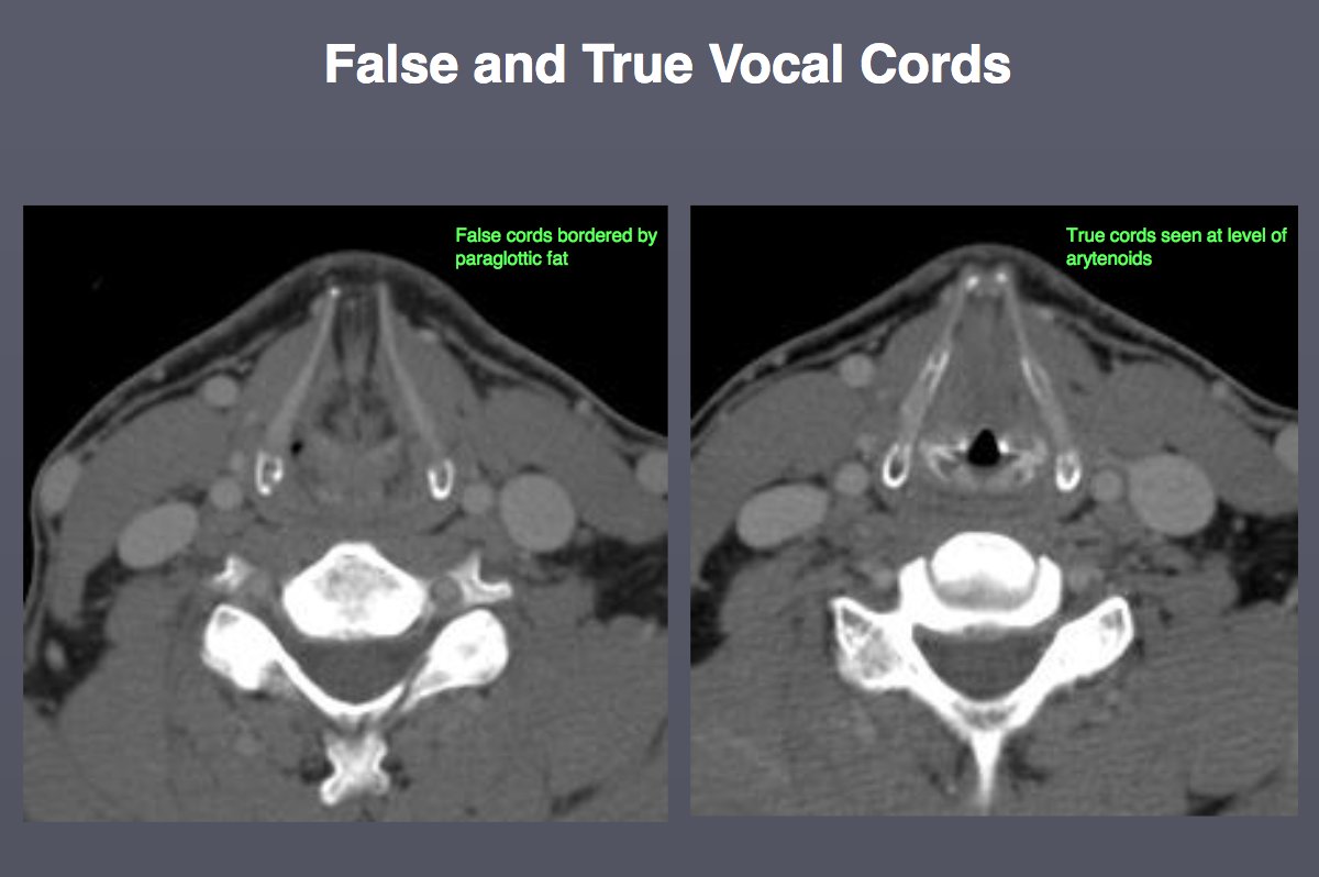
IU Radiology and Imaging Sciences on X: "RT @JeffersonRads: #HNRad anatomy  pearl from Dr N Rao - False vocal cords (supraglottic) ➡ bordered by  paraglottic fat True vocal cords (g…" / X
IU Radiology and Imaging Sciences on X: "RT @JeffersonRads: #HNRad anatomy pearl from Dr N Rao - False vocal cords (supraglottic) ➡ bordered by paraglottic fat True vocal cords (g…" / X
Larynx | Radiology Key

Larynx and Hypopharynx | Radiology Key

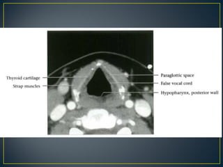
Larynx anatomy CT and MRI | PPT

IU Radiology and Imaging Sciences on X: "RT @JeffersonRads: #HNRad anatomy  pearl from Dr N Rao - False vocal cords (supraglottic) ➡ bordered by  paraglottic fat True vocal cords (g…" / X
IU Radiology and Imaging Sciences on X: "RT @JeffersonRads: #HNRad anatomy pearl from Dr N Rao - False vocal cords (supraglottic) ➡ bordered by paraglottic fat True vocal cords (g…" / X

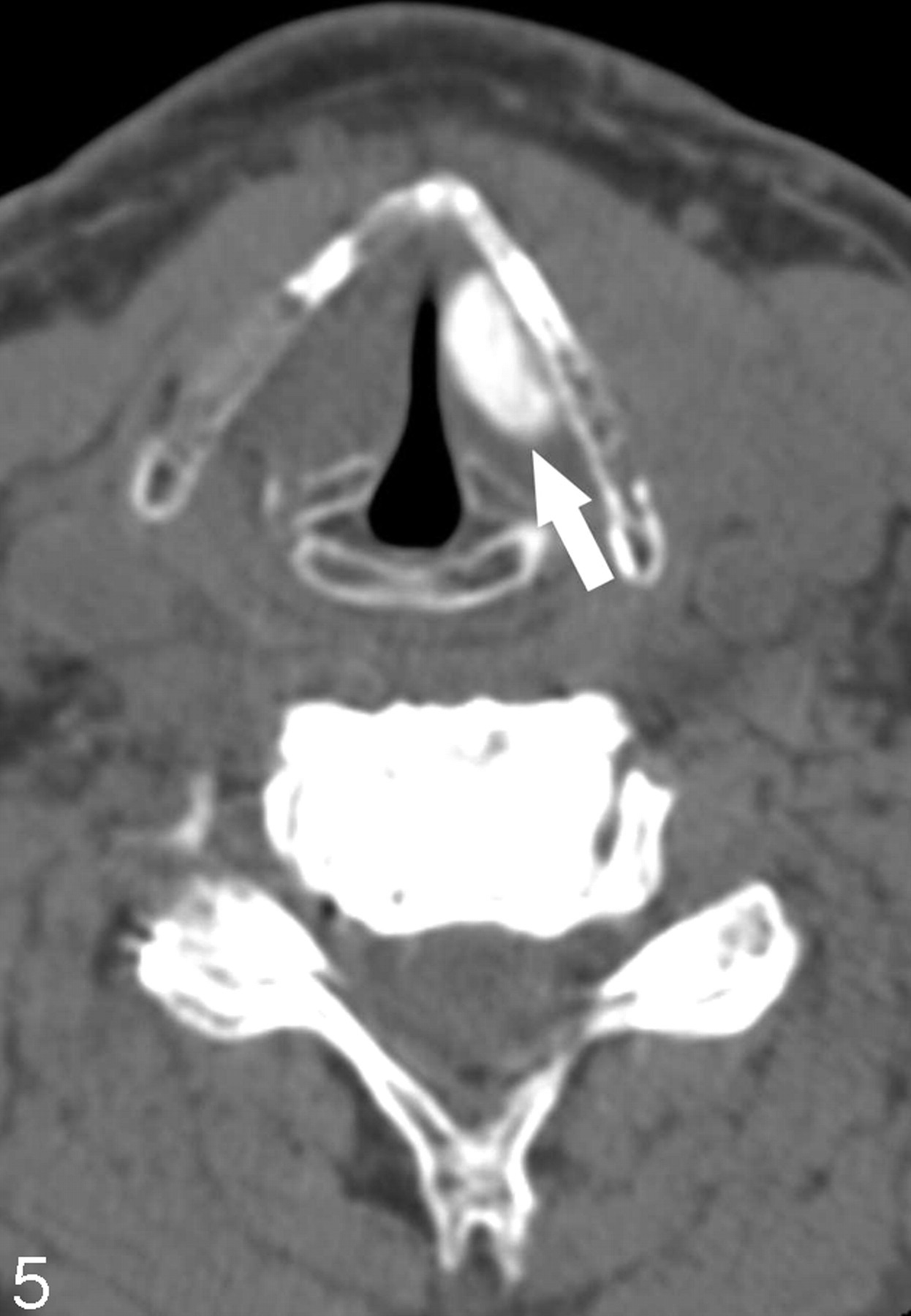
CT Assessment of Vocal Cord Medialization | American Journal of  Neuroradiology
CT Assessment of Vocal Cord Medialization | American Journal of Neuroradiology

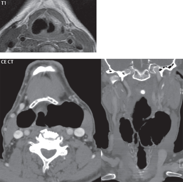
MRI T1 weighted image showing hyper-intense lesion in the right false... |  Download Scientific Diagram
MRI T1 weighted image showing hyper-intense lesion in the right false... | Download Scientific Diagram

Larynx | Radiology Key

Presentation1.pptx, radiological imaging of the larngeal diseases. | PPT

Full Story larynx Imaging CT MRI Dr Ahmed Esawy | PPT

CT Assessment of Vocal Cord Medialization | American Journal of  Neuroradiology
CT Assessment of Vocal Cord Medialization | American Journal of Neuroradiology